Thyroid and Parathyroid Surgery

Skull Base Surgery

Robotic and Laser Surgery

reconstructive surgery

Oral Cancer Surgery

Head & Neck Oncology Surgery

Gestational Trophoblastic Tumor Treatment
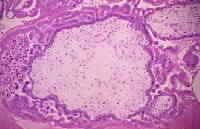
Gestational trophoblastic disease (GTD) can be benign or malignant. Histologically, it is classified into hydatidiform mole, invasive mole (chorioadenoma destruens), choriocarcinoma, and placental site trophoblastic tumor (PSTT). Those that invade locally or metastasize are collectively known as gestational trophoblastic neoplasia (GTN). Hydatidiform mole is the most common form of GTN. While invasive mole and choriocarcinoma are malignant, a hydatidiform mole can behave in a malignant or benign fashion .Histologic section of a complete hydatidiform mol...Histologic section of a complete hydatidiform mole stained with hematoxylin and eosin. Villi of different sizes are present. The large villous in the center exhibits marked edema with a fluid-filled central cavity known as cisterna. Marked proliferation of the trophoblasts is observed. The syncytiotrophoblasts stain purple, while the cytotrophoblasts have a clear cytoplasm and bizarre nuclei. No fetal blood vessels are in the mesenchyme of the villi. No methods exist to accurately predict the clinical behavior of a hydatidiform mole by histopathology. The clinical course is defined by the patient's serum human chorionic gonadotropin (hCG) curve after evacuation of the mole. In 80% of patients with a benign hydatidiform mole, serum hCG levels steadily drop to normal within 8-12 weeks after evacuation of the molar pregnancy. In the other 20% of patients with a malignant hydatidiform mole, serum hCG levels either rise or plateau.1,2
...more
Intraocular Melanoma Treatment
If the doctor suspects that a spot on the skin is melanoma, the patient will need to have a biopsy. A biopsy is the only way to make a definite diagnosis. In this procedure, the doctor tries to remove all of the suspicious-looking growth. This is an excisional biopsy. If the growth is too large to be removed entirely, the doctor removes a sample of the tissue. The doctor will never "shave off" or cauterize a growth that might be melanoma. A biopsy can usually be done in the doctor’s office using local anesthesia. A pathologist then examines the tissue under a microscope to check for cancer cells. Sometimes it is helpful for more than one pathologist to check the tissue for cancer cells.
...more
Bile Duct Treatment India
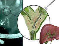
A bile duct is any of a number of long tube-like structures that carry bile. Bile, required for the digestion of food, is excreted by the liver into passages that carry bile toward the hepatic duct, which joins with the cystic duct (carrying bile to and from the gallbladder) to form the common bile duct, which opens into the intestine. The biliary tree (see below) is the whole network of various sized ducts branching through the liver. The path is as follows: Bile canaliculi Canals of Hering interlobular bile ducts intrahepatic bile ducts left and right hepatic ducts merge to form common hepatic duct exits liver and joins cystic duct (from gall bladder) forming common bile duct joins with pancreatic duct forming ampulla of Vater enters duodenum
...more
Basal Cell Carcinoma Treatment
If you have a change on the skin, the doctor must find out whether it is due to cancer or to some other cause. Your doctor removes all or part of the area that does not look normal. The sample goes to a lab. A pathologist checks the sample under a microscope. This is a biopsy. A biopsy is the only sure way to diagnose skin cancer. You may have the biopsy in a doctor's office or as an outpatient in a clinic or hospital. Where it is done depends on the size and place of the abnormal area on your skin. You probably will have local anesthesia. There are four common types of skin biopsies: Punch biopsy: The doctor uses a sharp, hollow tool to remove a circle of tissue from the abnormal area. Incisional biopsy: The doctor uses a scalpel to remove part of the growth. Excisional biopsy: The doctor uses a scalpel to remove the entire growth and some tissue around it. Shave biopsy: The doctor uses a thin, sharp blade to shave off the abnormal growth.
...more
Appendix Cancer Treatment
The appendix is a small hollow tube attached to the large colon (the large colon is also called large bowel or large intestine). The appendix is approximately 4 inches long and shaped like a worm. The appendix serves no known purpose, although it is thought to possibly play a role in the immune system. Very rarely, the appendix may become cancerous. Since the appendix is attached to the colon, appendix cancer is considered a type of colorectal cancer. Colorectal cancers are also part of a larger group of cancers of gastrointestinal tract, or GI cancers. Cancer of the appendix may cause appendicitis or cause the appendix to rupture. Sometimes this is the first symptom of appendix cancer. A ruptured appendix may cause a very serious condition called peritonitis, which is an infection of the lining of the abdomen and pelvis. A cancerous tumor of the appendix may also "seed" the abdomen with cancer cells. This may cause more cancerous tumors to develop in the abdomen before it is discovered. See Peritoneal Surface Malignancies and Peritoneal Carcinomatosis. Many times there are no symptoms of appendix cancer until it has progressed and is advanced. Abdominal discomfort and bloating of the abdomen can be signs of advanced appendiceal cancer There are five most common (though still VERY rare) varieties of appendix cancer: Malignant Carcinoid, Mucinous Adenocarcinoma, Adenocarcinoma, Adenocarcinoid, and Signet Ring Adenocarcinoma. To learn more about these varieties of appendix cancer, treatment and prognosis, click the links here or on the left side of this screen.
...more
Adrenocortical Carcinoma Treatment (Adult)
Adrenocortical carcinoma, also adrenal cortical carcinoma (ACC) and adrenal cortex cancer, is an aggressive cancer originating in the cortex (steroid hormone-producing tissue) of the adrenal gland. Adrenocortical carcinoma is a rare tumor, with incidence of 1-2 per million population annually. Adrenocortical carcinoma has a bimodal distribution by age, with cases clustering in children under 6, and in adults 30-40 years old. Adenocortical carcinoma is remarkable for the many hormonal syndromes which can occur in patients with steroid hormone-producing ("functional") tumors, including Cushing's syndrome, Conn syndrome, virilization, and feminization. Adrenocortical carcinoma has often invaded nearby tissues or metastasized to distant organs at the time of diagnosis, and the overall 5-year survival rate is only 20-35%. Laboratory findings Hormonal syndromes should be confirmed with laboratory testing. Laboratory findings in Cushing syndrome include increased serum glucose (blood sugar) and increased urine cortisol. Adrenal virilism is confirmed by the finding of an excess of serum androstenedione and dehydroepiandrosterone. Findings in Conn syndrome include low serum potassium, low plasma renin activity, and high serum aldosterone. Feminization is confirmed with the finding of excess serum estrogen Radiology Radiological studies of the abdomen, such as CT scans and magnetic resonance imaging are useful for identifying the site of the tumor, differentiating it from other diseases, such as adrenocortical adenoma, and determining the extent of invasion of the tumor into surrounding organs and tissues. CT scans of the chest and bone scans are routinely performed to look for metastases to the lungs and bones respectively. These studies are critical in determining whether or not the tumor can be surgically removed, the only potential cure at this time.
...more
Acute Myeloid Leukemia Treatment (Adult)
This page has important information about leukemia,* cancer that starts in blood cells. Each year, leukemia is diagnosed in about 29,000 adults and 2,000 children in the United States. This discusses possible causes, symptoms, diagnosis, treatment, and follow up care. It also has information to help people with leukemia and their families cope with the disease. Research is increasing what we know about leukemia. Scientists are studying its causes. They are also finding better ways to treat this disease. Because of research, adults and children with leukemia can look forward to a better quality of life and less chance of dying from the disease. What Is Leukemia cancer? Leukemia is a type of cancer. Cancer is a group of many related diseases. All cancers begin in cells, which make up blood and other tissues. Normally, cells grow and divide to form new cells as the body needs them. When cells grow old, they die, and new cells take their place. Sometimes this orderly process goes wrong. New cells form when the body does not need them, and old cells do not die when they should. Leukemia is cancer that begins in blood cells.
...more
Acute Lymphocytic Leukemia Treatment (Adult)
Treatment and Diagnosis: Acute Lymphoblastic Leukemia If a person has symptoms that suggest leukemia, the doctor may do a physical exam and ask about the patient's personal and family medical history. The doctor also may order laboratory tests, especially blood tests. The exams and tests may include the following: Physical exam — The doctor checks for swelling of the lymph nodes, spleen, and liver. Blood tests — The lab checks the level of blood cells. Leukemia causes a very high level of white blood cells. It also causes low levels of platelets and hemoglobin, which is found inside red blood cells. The lab also may check the blood for signs that leukemia has affected the liver and kidneys. Biopsy — The doctor removes some bone marrow from the hipbone or another large bone. A pathologist examines the sample under a microscope. The removal of tissue to look for cancer cells is called a biopsy. A biopsy is the only sure way to know whether leukemia cells are in the bone marrow. There are two ways the doctor can obtain bone marrow. Some patients will have both procedures: Bone marrow aspiration: The doctor uses a needle to remove samples of bone marrow. Bone marrow biopsy: The doctor uses a very thick needle to remove a small piece of bone and bone marrow. Local anesthesia helps to make the patient more comfortable. Cytogenetics — The lab looks at the chromosomes of cells from samples of peripheral blood, bone marrow, or lymph nodes. Spinal tap — The doctor removes some of the cerebrospinal fluid (the fluid that fills the spaces in and around the brain and spinal cord). The doctor uses a long, thin needle to remove fluid from the spinal column. The procedure takes about 30 minutes and is performed with local anesthesia. The patient must lie flat for several hours afterward to keep from getting a headache. The lab checks the fluid for leukemia cells or other signs of problems. Chest x-ray — The x-ray can reveal signs of disease in the chest.
...more
Complex Reconstruction of head and neck defects

Robotic and Laser Surgery

head cancer doctor consultant, throat cancer doctor consultant

Acute Lymphocytic Leukemia Treatment (Childhood)
Acute Lymphoblastic Leukemia, Adult Treatment and Diagnosis: If a person has symptoms that suggest leukemia, the doctor may do a physical exam and ask about the patient's personal and family medical history. The doctor also may order laboratory tests, especially blood tests. The exams and tests may include the following: Physical exam — The doctor checks for swelling of the lymph nodes, spleen, and liver. Blood tests — The lab checks the level of blood cells. Leukemia causes a very high level of white blood cells. It also causes low levels of platelets and hemoglobin, which is found inside red blood cells. The lab also may check the blood for signs that leukemia has affected the liver and kidneys. Biopsy — The doctor removes some bone marrow from the hipbone or another large bone. A pathologist examines the sample under a microscope. The removal of tissue to look for cancer cells is called a biopsy. A biopsy is the only sure way to know whether leukemia cells are in the bone marrow. There are two ways the doctor can obtain bone marrow. Some patients will have both procedures: Bone marrow aspiration: The doctor uses a needle to remove samples of bone marrow. Bone marrow biopsy: The doctor uses a very thick needle to remove a small piece of bone and bone marrow. Local anesthesia helps to make the patient more comfortable. Cytogenetics — The lab looks at the chromosomes of cells from samples of peripheral blood, bone marrow, or lymph nodes. Spinal tap — The doctor removes some of the cerebrospinal fluid (the fluid that fills the spaces in and around the brain and spinal cord). The doctor uses a long, thin needle to remove fluid from the spinal column. The procedure takes about 30 minutes and is performed with local anesthesia. The patient must lie flat for several hours afterward to keep from getting a headache. The lab checks the fluid for leukemia cells or other signs of problems. Chest x-ray — The x-ray can reveal signs of disease in the chest.
...more
Acute Myeloid Leukemia Treatment (Childhood)
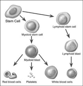
This page has important information about leukemia,* cancer that starts in blood cells. Each year, leukemia is diagnosed in about 29,000 adults and 2,000 children in the United States. This discusses possible causes, symptoms, diagnosis, treatment, and follow up care. It also has information to help people with leukemia and their families cope with the disease. Research is increasing what we know about leukemia. Scientists are studying its causes. They are also finding better ways to treat this disease. Because of research, adults and children with leukemia can look forward to a better quality of life and less chance of dying from the disease. What Is Leukemia cancer? Leukemia is a type of cancer. Cancer is a group of many related diseases. All cancers begin in cells, which make up blood and other tissues. Normally, cells grow and divide to form new cells as the body needs them. When cells grow old, they die, and new cells take their place. Sometimes this orderly process goes wrong. New cells form when the body does not need them, and old cells do not die when they should. Leukemia is cancer that begins in blood cells. Normal Blood Cells Blood cells form in the bone marrow. Bone marrow is the soft material in the center of most bones. Immature blood cells are called stem cells and blasts. Most blood cells mature in the bone marrow and then move into the blood vessels. Blood flowing through the blood vessels and heart is called the peripheral blood.
...more
AIDS-Related Cancer Treatment
After AIDS-related lymphoma has been diagnosed, tests are done to find out if cancer cells have spread within the lymph system or to other parts of the body. The process used to find out if cancer cells have spread within the lymph system or to other parts of the body is called staging. The information gathered from the staging process determines the stage of the disease. It is important to know the stage in order to plan treatment, but AIDS-related lymphoma is usually advanced when it is diagnosed. The following tests and procedures may be used in the staging process: CT scan (CAT scan): A procedure that makes a series of detailed pictures of areas inside the body, taken from different angles. The pictures are made by a computer linked to an x-ray machine. A dye may be injected into a vein or swallowed to help the organs or tissues show up more clearly. This procedure is also called computed tomography, computerized tomography, or computerized axial tomography. PET scan (positron emission tomography scan): A procedure to find malignant tumor cells in the body. A small amount of radioactive glucose (sugar) is injected into a vein. The PET scanner rotates around the body and makes a picture of where glucose is being used in the body. Malignant tumor cells show up brighter in the picture because they are more active and take up more glucose than normal cells do. MRI (magnetic resonance imaging): A procedure that uses a magnet, radio waves, and a computer to make a series of detailed pictures of areas inside the body. A substance called gadolinium is injected into the patient through a vein. The gadolinium collects around the cancer cells so they show up brighter in the picture. This procedure is also called nuclear magnetic resonance imaging (NMRI).
...more
AIDS-Related Lymphoma Treatment
Individuals infected with human immunodeficiency virus (HIV) have A high risk of developing lymphomas. Approximately 4% of people with acquired immunodeficiency syndrome (AIDS) have non-Hodgkin Lymphoma (NHL) at diagnosis and at least the same proportion develop NHL in others during the course of illness.1 AIDS-related lymphoma (ARL) can be divided into 3 types on the basis of areas of involvement: Systemic NHL Primary central nervous system lymphoma (PCNSL) Primary effusion lymphomas ("body cavity lymphoma") Systemic NHL is the most common variety of AIDS-related lymphoma (ARL), followed by PCNSL, which is less common but not rare, and primary effusion lymphoma, which is a rare disease. Histologically, the most common variants are diffuse, large B-cell lymphoma and small, noncleaved cell lymphoma, including Burkitt and/or Burkitt-like lymphoma (see Images 1-4 or below)
...more
Anal Cancer Treatment
Anal Cancer Treatment

Astrocytoma, Childhood Cerebellar Treatment
Treatment and Diagnosis: If a person has symptoms that suggest a brain tumor, the doctor may perform one or more of the following procedures: Physical exam — The doctor checks general signs of health. Neurologic exam — The doctor checks for alertness, muscle strength, coordination, reflexes, and response to pain. The doctor also examines the eyes to look for swelling caused by a tumor pressing on the nerve that connects the eye and brain. CT scan — An x-ray machine linked to a computer takes a series of detailed pictures of the head. The patient may receive an injection of a special dye so the brain shows up clearly in the pictures. The pictures can show tumors in the brain. MRI — A powerful magnet linked to a computer makes detailed pictures of areas inside the body. These pictures are viewed on a monitor and can also be printed. Sometimes a special dye is injected to help show differences in the tissues of the brain. The pictures can show a tumor or other problem in the brain. The doctor may ask for other tests: Angiogram — Dye injected into the bloodstream flows into the blood vessels in the brain to make them show up on an x-ray. If a tumor is present, the doctor may be able to see it on the x-ray. Skull x-ray — Some types of brain tumors cause calcium deposits in the brain or changes in the bones of the skull. With an x-ray, the doctor can check for these changes. Spinal tap — The doctor may remove a sample of cerebrospinal fluid (the fluid that fills the spaces in and around the brain and spinal cord). This procedure is performed with local anesthesia. The doctor uses a long, thin needle to remove fluid from the spinal column. A spinal tap takes about 30 minutes. The patient must lie flat for several hours afterward to keep from getting a headache. A laboratory checks the fluid for cancer cells or other signs of problems.
...more
Astrocytoma, Childhood Cerebral Treatment
Treatment and Diagnosis: If a person has symptoms that suggest a brain tumor, the doctor may perform one or more of the following procedures: Physical exam — The doctor checks general signs of health. Neurologic exam — The doctor checks for alertness, muscle strength, coordination, reflexes, and response to pain. The doctor also examines the eyes to look for swelling caused by a tumor pressing on the nerve that connects the eye and brain. CT scan — An x-ray machine linked to a computer takes a series of detailed pictures of the head. The patient may receive an injection of a special dye so the brain shows up clearly in the pictures. The pictures can show tumors in the brain. MRI — A powerful magnet linked to a computer makes detailed pictures of areas inside the body. These pictures are viewed on a monitor and can also be printed. Sometimes a special dye is injected to help show differences in the tissues of the brain. The pictures can show a tumor or other problem in the brain. The doctor may ask for other tests: Angiogram — Dye injected into the bloodstream flows into the blood vessels in the brain to make them show up on an x-ray. If a tumor is present, the doctor may be able to see it on the x-ray. Skull x-ray — Some types of brain tumors cause calcium deposits in the brain or changes in the bones of the skull. With an x-ray, the doctor can check for these changes.
...more
Bladder Cancer Treatment
Treatment and Diagnosis: If a patient has symptoms that suggest bladder cancer, the doctor may check general signs of health and may order lab tests. The person may have one or more of the following procedures: Physical exam -- The doctor feels the abdomen and pelvis for tumors. The physical exam may include a rectal or vaginal exam. Urine tests -- The laboratory checks the urine for blood, cancer cells, and other signs of disease. Intravenous pyelogram -- The doctor injects dye into a blood vessel. The dye collects in the urine, making the bladder show up on x-rays. Cystoscopy -- The doctor uses a thin, lighted tube (cystoscope) to look directly into the bladder. The doctor inserts the cystoscope into the bladder through the urethra to examine the lining of the bladder. The patient may need anesthesia for this procedure The doctor can remove samples of tissue with the cystoscope. A pathologist then examines the tissue under a microscope. The removal of tissue to look for cancer cells is called a biopsy. In many cases, a biopsy is the only sure way to tell whether cancer is present. For a small number of patients, the doctor removes the entire cancerous area during the biopsy. For these patients, bladder cancer is diagnosed and treated in a single procedure.
...more
Osteosarcoma Treatment
Bone Cancer, Osteosarcoma/Malignant Fibrous Histiocytoma Bone tumor is an inexact term, which can be used for both benign and malignant abnormal growths found in bone, but is most commonly used for primary tumors of bone, such as osteosarcoma. It is less exactly applied to secondary, or metastatic tumors found in bone. Bone tumors may be classified as "primary tumors" which originate in the bone, and "secondary tumors" which originate elsewhere. Primary tumors of bone can be divided into benign tumors and cancers. Common benign bone tumors may be neoplastic, developmental, traumatic, infectious, or inflammatory in etiology. Examples of benign bone tumors include osteoma, osteochondroma, aneurysmal bone cyst, and fibrous dysplasia. Malignant primary bone tumors include osteosarcoma, chondrosarcoma, Ewing's sarcoma, malignant fibrous histiocytoma, fibrosarcoma, and other sarcoma types. Multiple myeloma is a hematologic cancer which also frequently presents as one or more bone tumors. The tailbone is a common location for a teratoma, known as a sacrococcygeal teratoma, and related germ cell tumors. Secondary bone tumors include metastatic tumors which have spread from other organs, such as the breast, lung, and prostate. Metastatic tumors more frequently involve the axial skeleton than the appendicular skeleton. Tumors which originate in the soft tissues may also secondarily involve bones through direct invasion.
...more
Brain Stem Glioma Treatment (Childhood)
Treatment and Diagnosis: If a person has symptoms that suggest a brain tumor, the doctor may perform one or more of the following procedures: Physical exam — The doctor checks general signs of health. Neurologic exam — The doctor checks for alertness, muscle strength, coordination, reflexes, and response to pain. The doctor also examines the eyes to look for swelling caused by a tumor pressing on the nerve that connects the eye and brain. CT scan — An x-ray machine linked to a computer takes a series of detailed pictures of the head. The patient may receive an injection of a special dye so the brain shows up clearly in the pictures. The pictures can show tumors in the brain. MRI — A powerful magnet linked to a computer makes detailed pictures of areas inside the body. These pictures are viewed on a monitor and can also be printed. Sometimes a special dye is injected to help show differences in the tissues of the brain. The pictures can show a tumor or other problem in the brain. The doctor may ask for other tests: Angiogram — Dye injected into the bloodstream flows into the blood vessels in the brain to make them show up on an x-ray. If a tumor is present, the doctor may be able to see it on the x-ray. Skull x-ray — Some types of brain tumors cause calcium deposits in the brain or changes in the bones of the skull. With an x-ray, the doctor can check for these changes.
...more
Brain Stem Glioma Treatment (Adult)
Treatment and Diagnosis: If a person has symptoms that suggest a brain tumor, the doctor may perform one or more of the following procedures: Physical exam — The doctor checks general signs of health. Neurologic exam — The doctor checks for alertness, muscle strength, coordination, reflexes, and response to pain. The doctor also examines the eyes to look for swelling caused by a tumor pressing on the nerve that connects the eye and brain. CT scan — An x-ray machine linked to a computer takes a series of detailed pictures of the head. The patient may receive an injection of a special dye so the brain shows up clearly in the pictures. The pictures can show tumors in the brain. MRI — A powerful magnet linked to a computer makes detailed pictures of areas inside the body. These pictures are viewed on a monitor and can also be printed. Sometimes a special dye is injected to help show differences in the tissues of the brain. The pictures can show a tumor or other problem in the brain.
...more
Brain Tumor Treatment (Childhood)
Brain Tumor, Central Nervous System Embryonal Tumors, Childhood Treatment and Diagnosis: If a person has symptoms that suggest a brain tumor, the doctor may perform one or more of the following procedures: Physical exam — The doctor checks general signs of health. Neurologic exam — The doctor checks for alertness, muscle strength, coordination, reflexes, and response to pain. The doctor also examines the eyes to look for swelling caused by a tumor pressing on the nerve that connects the eye and brain. CT scan — An x-ray machine linked to a computer takes a series of detailed pictures of the head. The patient may receive an injection of a special dye so the brain shows up clearly in the pictures. The pictures can show tumors in the brain. MRI — A powerful magnet linked to a computer makes detailed pictures of areas inside the body. These pictures are viewed on a monitor and can also be printed. Sometimes a special dye is injected to help show differences in the tissues of the brain. The pictures can show a tumor or other problem in the brain.
...more
brain tumor treatment service
Treatment and Diagnosis: If a person has symptoms that suggest a brain tumor, the doctor may perform one or more of the following procedures: Physical exam — The doctor checks general signs of health. Neurologic exam — The doctor checks for alertness, muscle strength, coordination, reflexes, and response to pain. The doctor also examines the eyes to look for swelling caused by a tumor pressing on the nerve that connects the eye and brain. CT scan — An x-ray machine linked to a computer takes a series of detailed pictures of the head. The patient may receive an injection of a special dye so the brain shows up clearly in the pictures. The pictures can show tumors in the brain. MRI — A powerful magnet linked to a computer makes detailed pictures of areas inside the body. These pictures are viewed on a monitor and can also be printed. Sometimes a special dye is injected to help show differences in the tissues of the brain. The pictures can show a tumor or other problem in the brain.
...moreBe first to Rate
Rate ThisOpening Hours